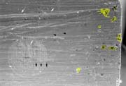
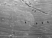
 SEM surface morphology of harmonica reeds.
The SEM, a.k.a., Scanning Electron Microscope, belongs to the family of the electron microscopes. You know those fancy and big instruments which scientists use to look the molecular world. Its characteristics include 3-D imaging with high resolution and depth of focus. I started working with SEM in the biological field when first entered the University of Bologna in 1983. My first original scientific contributions can be found on the journal of Scanning Electron Microscopy, Chicago and Cells and Materials, Chicago. I had also the privilege to be a reviewer for those journals. I also wrote a couple of books, in italian, on this technical subject. Then I started to be a full time bread and butter pathologist, working with the more conventional light microscope, and in the spare time an avid harmonica player. Now I have the opportunity to use the SEM again: only few minutes a week. These new attempts are directed to satisfy my current curiosity. How is the surface structure of a reed ? Well I punched out a bad working reed, I covered it with a thin layer of gold (approximately 30 nm thick) under vacuum and finally I observed it under the SEM. To the best of my knowledge these are the first SEM images of a harmonica reed (Hohner brand). My aim is to go on this research hoping in the help of the harp comunity.
